VENEPUNCTURE
venepuncture resource file
"Venepuncture is the term used for the procedure of entering a vein with a needle"
(Rowland 1991)
|
 |
|
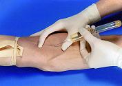
Venepuncture is a procedure performed to obtain venous blood for analysis. The various blood chemistry and haematology tests offered by most laboratories represent an economical way by which a great amount of information about a patients physical condition, at the time of the testing can be made available to the Physician. Most blood tests fall within one of two categories: screening or diagnostic. Screening tests are used to detect a disease when there is little or no evidence that a person has a suspected disease. Diagnostic tests are utilised when a specific disease is suspected, in order to verify its presence and the severity of the disease. It must be taken into account when using these results though that no blood result is completely accurate all of the time. Commonly carried out previously by junior doctors, it is more often, now performed by nurses (Inwood 1996), due to the increased freedom which has been provided within The Scope of Professional Practice, Phlebotomists or other suitably trained person. What becomes clear, is that by being trained in the art of venepuncture,it is yet another procedure for which we become accountable for (NMC 2002). What we need to be aware of is that by carrying out the procedure incorrectly, not only can it have a damaging effect on the individual, but it can be classed in legal terms as "assault" (McConnell and Mackay 1996). Problems arise when the first attempt proves unsuccessful and the professional then goes on to attempt the procedure a second time without gaining further consent from the patient. Each time a venepuncture is attempted, the practitioner must ensure that they obtain the patients consent.
|
 |
WHICH VEINS SHOULD BE USED
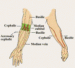
Which veins should and should not be usedThe usual choice of veins which are most suited are the superficial veins of the upper limb, namely the Basilic Vein, the Cephalic Vein, the Median Cubital Vein or the veins in the dorsal aspect of the hand. These veins are numerous and accessible ensuring that the procedure can be performed safely and with the minimum of discomfort.
VEINS WHICH ARE FIBROSED, FRAGILE OR INFLAMMED SHOULD BE AVOIDED.
STRUCTURE OF VEINS
Structure of the VeinsVeins are vessels that carry blood back from the heart. They have thinner walls than that of an artery, due to the blood flowing at a lower pressure. Veins consist of 3 layers:
The Tunica Interna, the inner layer, is a smooth endothelial lining which allows the passage of blood cells. Within this layer are folds of endothelium called valves, which keeps blood moving towards the heart by preventing back flow.
The Tunica Media, the middle layer, is composed of muscular tissue and nerve Fibres, both vaso-constrictors and vaso-dilators, which can stimulate the vein to both contract or relax.
The Tunica Externa is the outer layer and consists of connective tissue which surrounds and supports the vessel
TYPES OF VEINS
Types of Veins
PROMINENT VEINS
Every Phlebotomists dream, large prominent and bouncy. Easy to see difficult to miss, but care should be taken not to insert the needle too far. Always run the needle along the vein, never through it.
DEEP VEINS
Palpitation of the veins is a skill that comes with practice. It is often the case that good veins lie just below the skins surface, and this is when you need to be guided by what you can feel rather than by what you can see.
THREADY VEINS.
These veins, once punctured, often bleed profusely, so extra care should be taken to ensure haemostasis following the procedure. They will also flatten out and become useless is a too severe vacuum is exerted during the procedure.
SUPERFICIAL VEINS.
Tiny veins which appear on the surface of the arm, particularly in the Elderly, and are not generally suitable for venepuncture. In these cases the are needs to be palpated to feel for deeper, larger veins.
FLOATING VEINS
They can be deceptive and on the point of entry, they can move. Before inserting the needle, the vein needs to be anchored (about 3-5cms) below the intended puncture site with the thumb or forefinger. Once the needle is well in position, the retaining hand can be moved to support the syringe/vacutainer.
THROMBOSED VEINS.
These veins have been subjected to overuse (either by continual venepuncture, drug abuse etc) and can be very difficult to obtain a sample from. They are hard and knotty to the touch.
VERY DEEP VEINS
Often associated with severely obese patients. Palpation in the regular sites can be used to locate a more suitable vein.


